Glossary
Contraction force measured at the greatest level of muscle activation that can be elicited under voluntary conditions (maximal voluntary effort).The EMG signal is often normalized with respect to the EMG recorded at MVC. Some individuals may not be able to fully activate a muscle during MVC and additional muscle force can be generated through electrical stimulation of the motor nerve supplying the muscle (twitch interpolation).
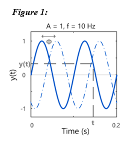
(McManus et al., 2021)The average number of action potentials discharged per second by a single motor unit.Typically measured in discharges per second, pulses per second (PPS) or Hertz (Hz). Hertz is a unit that measures the number of cycles per second of a periodic, sinusoidal waveform and is thus not strictly applicable to the measurement of aperiodic motor unit firing.
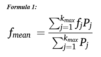
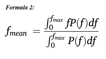
(McManus et al., 2021)Defines the centroid or barycenter of the power spectrum of the signal that is the frequency around which the frequency components of the spectrum are grouped. Mathematically defined as the normalised, one-sided, first-order spectral moment of the EMG power spectral density. The mean frequency (or mean power frequency), fmean, is defined in Formula 1. Pj is the power of the jth frequency component of the EMG power spectrum and kmax is the index of the highest frequency component present in the signal (the highest frequency is usually taken as half of the sampling frequency). Related terms: Median spectral frequency, power spectrum, Fourier transform, frequency. MNF is usually higher than the median spectral frequency because of the skewed shape of the EMG power spectrum. The equation for fmean can also be expressed in continuous time as in Formula 2. Fmax is the highest frequency component present in the signal.
(McManus et al., 2021)

The frequency which divides the EMG power spectrum into two regions, each containing half of the total power within the signal epoch. It is also defined as the 50th percentile of the power spectrum, which means that the frequency components below it contain 50% of the total power and those above it contain 50% of the total power. The median frequency, fmed, is defined as in Formula 1. Pj is the power of the jth frequency component of the EMG power spectrum, kfmed is the index of the median frequency and kmax is the index of the highest frequency component present in the signal (the highest frequency is usually half of the sampling frequency). Note that fmed = Δf·kfmed, where Δf is the frequency resolution. Related terms: Mean spectral frequency, power spectrum, Fourier transform, frequency. MDF and mean spectral frequency have similar properties. The equation for fmed can also be expressed in continuous time, as shown in Formula 2. Fmax is the highest frequency component present in the signal. The value fmax is often replaced by half of the sampling frequency (fs/2) since P(f) = 0 at frequencies greater than fmax.
(McManus et al., 2021)


Unwanted voltage fluctuations present in the EMG signal due to movement between the electrode and tissue or the movement of cables connected to the recording electrodes. Motion artefact at the electrode-tissue interface encompasses both movement of the electrode and the underlying skin and deformation or stretching of the skin underneath the electrode. It can be reduced by careful skin preparation and secure placement of the electrodes. Most of the power in EMG signals occurring due to motion artefact will lie below 20 Hz and can be attenuated by high-pass filtering the signal.
(McManus et al., 2021)Neurons that integrate information from sources within the nervous system and transmit information to the muscle fibers that they innervate. The alpha motoneuron cell body is located in the spinal cord, or brainstem, and the axon leaves these structures to innervate a group of muscle fibers. In the context of EMG, the term motoneuron typically refers to alpha motoneurons, which innervate extrafusal muscle fibers, rather than gamma motoneurons, which innervate smaller intrafusal fibers within the muscle spindle. Alpha motoneurons are known as the final common pathway in the processing of motor commands, as all activation signals from the brainstem/spinal cord to muscle are transmitted through them.
(McManus et al., 2021)A motor-evoked potential (MEP) is the electrical response recorded from a muscle following stimulation of the motor cortex, typically using transcranial magnetic stimulation (TMS) (Rossini et al., 1994). The MEP reflects the compound muscle action potential elicited by descending motor signals and is commonly recorded using surface EMG. MEPs are used to assess the excitability of the corticospinal pathway, and they are distinct from the mechanical twitch response (i.e., force) that may also follow cortical or peripheral stimulation. Note: Although TMS is the most common technique, MEPs can also be elicited using electrical stimulation of the cortex in some research or clinical settings.
(Rossini et al., 1994)A motoneuron and all of the muscle fibers it innervates.All muscle fibers belonging to a motor unit are activated every time the motoneuron discharges (in normal, healthy physiology). The muscle fibers of a motor unit (without the motoneuron) define the muscle unit.
(McManus et al., 2021)The summation of individual muscle fibre action potentials from all the fibers of a motor unit in response to a discharge of its motoneuron.The motor unit action potential detected by an electrode system will depend on the electrode configuration and the position/orientation of the electrode relative to the muscle fibers.
(McManus et al., 2021)Activation of a motor unit to participate in a muscle contraction. Related term: Recruitment threshold of a motor unit.During voluntary muscle contractions where there is a gradual increase in force, motor units are usually recruited in an orderly fashion referred to as “Henneman’s size principle”. This states that the threshold of motoneuron activation is determined by the size of the neuron, with recruitment occurring in approximate order of size –from the smallest motor units (typically innervated by the smallest motoneurons) to the largest (typically innervated by the largest motoneurons). It should be noted that the size principle may not apply to all types of muscle contractions (e.g., during ballistic or electrically stimulated muscle contractions).
(McManus et al., 2021)We value your feedback
Let us know how helpful you found the recommendations above and how we can improve: