Glossary
A method of recording EMG using three electrodes and three differential amplifiers, which is used to reduce interference and crosstalk in the EMG signal.EMG is recorded from three electrodes aligned over the active muscle (typically with equidistant spacing). The signals are passed into two differential amplifiers (with one input common to both amplifiers) to obtain the difference between the first and second signals, and between the second and third signals. The output signals from the two differential amplifiers (i.e., bipolar or single differential EMG signals) are then used as an input to a third differential amplifier to obtain the double differential signal. Performing the differential subtraction twice results in a higher attenuation of signals originating from muscles that are further away (as these signals will appear similar, i.e., as common-mode signals, at all amplifier inputs).
(McManus et al., 2021)Two successive action potentials of the same motor unit close together in time. Historically, the interspike interval for doublets has been defined as 2.5–20 ms, however, it may be more accurate to define a doublet as an interspike interval that is significantly shorter than the mean interspike interval for a given motoneuron. A doublet is also typically followed by a long interspike interval.
(McManus et al., 2021)A technique widely used in electrocardiography, and occasionally in electromyography, whereby the common-mode voltage (primarily due to power line interference) on the body is negatively fed back (i.e., the phase is reversed) to a third electrode, through a feedback amplifier, driving the common mode voltage to a lower level and thereby reducing the power line interference. The additional ECG electrode is typically applied to the right leg (hence, the name).This technique is occasionally used in surface electromyography where the additional feedback electrode is not necessarily applied to the right leg.
(McManus et al., 2021)Unwanted voltage fluctuations due to the ECG signal, typically present when EMG electrodes are applied to the chest, back or abdominal muscles. ECG interference can be attenuated by processing the EMG signal post-recording, as it may not be sufficiently reduced by differential detection or the DRL technique.
(McManus et al., 2021)Sensor made of conductive material that couples the electrical potentials generated within the body to an electronic device. In EMG, reference electrodes are typically placed over an electrically silent area such as a tendon or bone (surface electrodes) or on the cannula (intramuscular Electromyographic signal electrode). In monopolar recording configurations, the detected EMG signal is the potential difference between the recording electrode and the reference electrode.
(McManus et al., 2021)The opposition (impedance) to the flow of the electrical current, as a function of frequency, at the contact point between the electrode and the skin surface. Lower electrode impedance is generally desired.
(McManus et al., 2021)The contact region between the electrode and the skin surface.The change in the current carrier (from ions to electrons) at the interface between the electrode and the skin generates random noise, the amplitude of which will depend on the contact surface, the electrode used and the skin condition. The impedance at the electrode-tissue interface may be reduced by conductive gel.
(McManus et al., 2021)The time-varying electrical potential recorded within a muscle, in the surrounding tissue or at the skin surface during muscle activity. It is the summation of the extracellular action potentials generated by muscle fibers within the vicinity of the electrode.
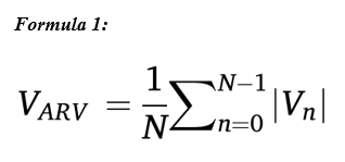
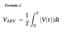
(McManus et al., 2021)An estimate of the average amplitude of a signal over a given time period. The ARV is defined as the average of the “rectified” (absolute value) signal over an epoch (or window) of duration T (or N samples). The average rectified value for one signal epoch, VARV, is defined as in Formula 1. N is the total number of samples being averaged (over the epoch of the EMG signal under analysis) and Vn is the value of the nth sample within the epoch. The epoch/window duration must be stated when reporting the VRMS. In practice, the mean value of the signal should be subtracted from the total signal length before rectifying. The epoch/window duration should also be stated when reporting VARV. The equation for VARV can also be expressed in continuous time, as shown in Formula 2. T is the length of the signal epoch in seconds.
(McManus et al., 2021)

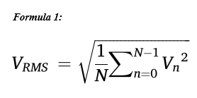
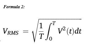
An estimate of the standard deviation (amplitude) of the signal over a given time window or epoch. The RMS value is the square root of the mean square value estimated over an epoch (or window) of duration T (or N samples). RMS is the square root of the power of the signal. The root mean square value, VRMS, for one signal epoch is defined in Formula 1, where N is the total number of samples being averaged (over the epoch of the EMG signal under analysis) and Vn is the value of the nth sample within the epoch. The epoch/window duration must be stated when reporting the VRMS. The equation for VRMS can also be expressed in continuous time as expressed in Formula 2, where T is the length of the signal epoch in seconds.
(McManus et al., 2021)

We value your feedback
Let us know how helpful you found the recommendations above and how we can improve: