Definition of the method
An M-wave is the compound muscle action potential evoked by electrical stimulation of the motoneurons innervating a muscle.
This method includes:
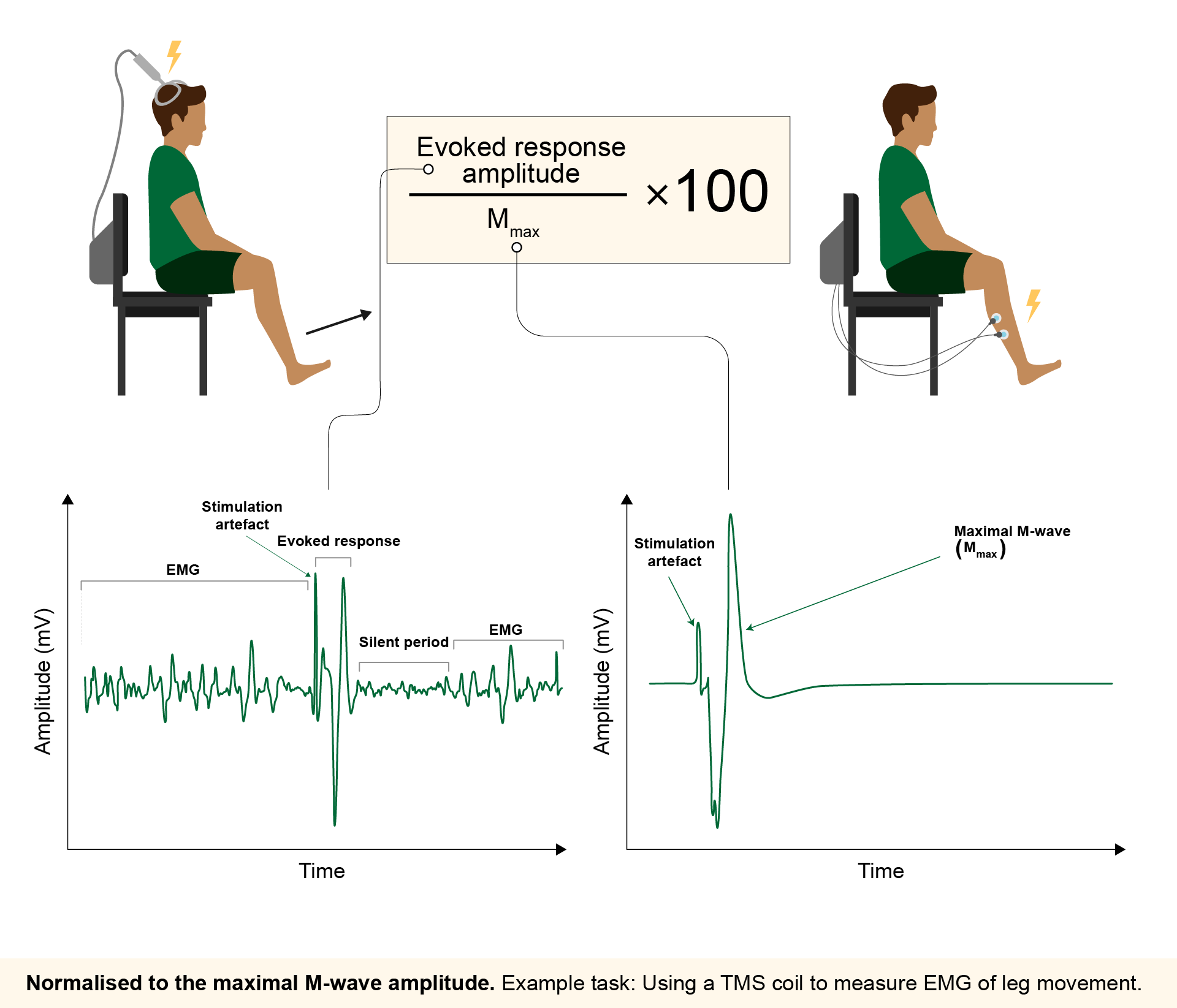
Several electrical and magnetic stimulation techniques are used to evoke compound muscle action potentials to study neurophysiological mechanisms (e.g., H-reflexes, F-waves, and motor-evoked potentials from transcranial magnetic stimulation of the motor cortex). The amplitudes of these evoked responses are often used to estimate the excitability status of the motoneuron pool, as these techniques recruit motoneurons via various neural circuits. However, changes in the amplitude of these evoked responses can result from both central (e.g., at the cortical level) and/or peripheral physiological processes (i.e., within the muscle). To remove the possible influence of peripheral mechanisms when interpreting these evoked responses, the amplitude from evoked responses should be normalised to the amplitude of the maximal compound muscle action potential (Mmax). The Mmax is a short-latency evoked response where all muscle fibres are presumably recruited via peripheral electrical stimulation of motor axons.
Let’s assume that we aim to estimate changes in corticospinal excitability by recording motor-evoked potentials (MEPs) before and after an intervention. Before the intervention, we record an MEP with amplitude Apre and an Mmax with amplitude Bpre. After the intervention, we record an MEP with amplitude Apost and an Mmax with amplitude Bpost. We then observe that both Apost and Bpost are smaller than their respective pre-intervention values. However, when comparing normalised values (i.e., Apre/Bpre vs Apost/Bpost), we find no differences between pre- and post-intervention. This suggests that the reduction in MEP amplitude was driven by a decrease in peripheral excitability rather than a change in the central mechanisms we aimed to study. Had we not normalised MEPs to Mmax, we might have mistakenly concluded that corticospinal excitability decreased following the intervention.
As the Mmax represents the maximal synchronised activation of motor units recorded by surface EMG electrodes, this normalisation method also allows researchers to estimate the proportion of a motor unit pool recruited by a stimulus (presented as a % Mmax). For example, eliciting a soleus H-reflex amplitude that is 10% of the soleus Mmax suggests that 10% of the soleus motoneurons are being recruited from the entire soleus motoneuron pool.